
Two Windows computers with software for image analysis.
Located in NRB 260F.
Structured illumination Zeiss Apotome confocal microscope
Commonly used fluorophores FITC (Alexa 488), CY2, CY3, rhodamine, Texas Red, CY5,
and DAPI.
Zeiss Zen software
MBF Bioscience Stereo Investigator system software provides the ability to make accurate, unbiased quantification of the number, length, area, and volume of cells, subcellular and macro structures in tissue specimens.
The Apotome is located in NRB room 260N

Used for cutting frozen tissues in preparation for microscopic analyses.
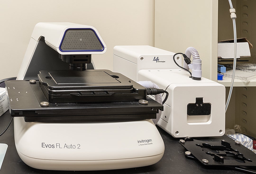
The EVOS FL Auto 2 image can be used for both brightfield and fluorescence microscopy of slides, well-plates, dishes, and flasks. The on stage incubator can control the temperature, humidity, and CO2 content, as well as create a hypoxia environment. The automation software can be used to acquire z-stacks, time lapse, and tiled images. It is equipped with 4X, 10X, and 20X objectives and DAPI, GFP, and Texas Red filter sets.
Evos is located in NRB room 355E.
Imaris’ software provides 3D/4D visualization and analysis tools, smart cell based segmentation, analysis on a per cell basis and discovery of intracellular relationships. The package includes automated tracking, detection of cell division and creation of interactive lineage trees along with statistical tests and a two-way interface for customization in Matlab, Java or Python.
The Imaris software is installed in a computer located in NRB room 260F.
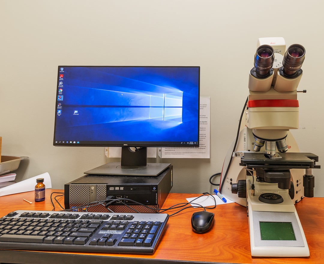
A Leica DMI4 upright microscope and a Leica DMI8 inverted microscope are available in NRB 440 for those who need a quick look at tissue samples. These are available on a walk in basis for authorized users.
For those needing longer time slots, other microscopes should be considered.
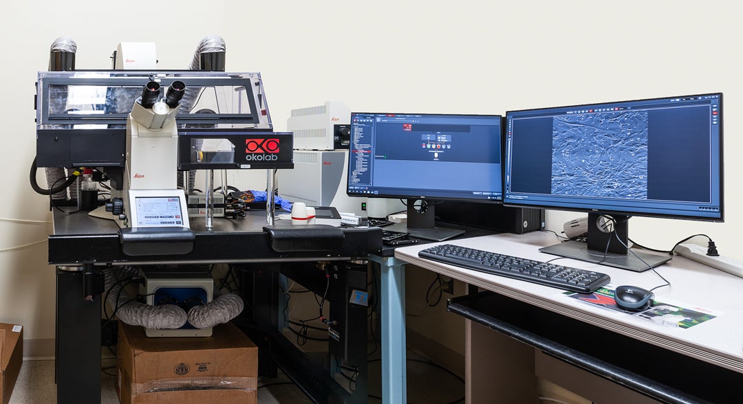
The Leica DMI8 inverted microscope is ideal for live imaging of cells in well plates. It is equipped with an automated stage, brightfield, and fluorescence (DAPI, GFP, Cy3, and Cy5 filters). It has a dry 10X and oil immersion 20X and 40X objectives.
IMPORTANT BIOSAFETY INFORMATION:
This instrument can be used at Biosafety Level 2, which allows for live imaging of human cells. Users must be trained and agree to follow all safety rules.
This system is located in NRB room 312.
Used to cut thin slices of material using a vibrating blade.
Located in NRB room 260N
ZEISS Laser Confocal Scanning Microscope with four diode lasers (405, 488, 561 and 640 nm), complete with three highly sensitive GaAsP detection and fast linear scanning.
Located in NRB 260S.


Samantha Butler, PhD
Co-Director

Peyman Golshani, MD, PhD
Co-Director


Quan Lin, PhD

Ronen Roshef, PhD
